

1Zhu C, 1Santana E, 1Urbano M, 1Quelhas A, 1Pereira R, 1Neves TR, 1Coimbra É, 1Bilhim T.
1Radiologia de Intervenção, Hospital Curry Cabral, Unidade Local de Saúde de São José
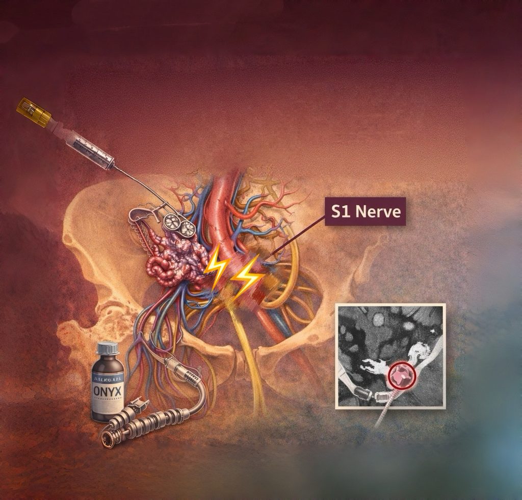
Illustration generated with AI using ChatGPT, based on the author’s prompt; final edits/corrections by the authors.
Clinical History
A 43-year-old male with a complex pelvic arteriovenous malformation (AVM), underwent embolization on April 2025, with placement of plugs in the anterior and posterior branches of the left internal iliac vein.
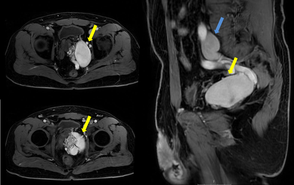
Figure 1 and Video1– Baseline pelvic AVM (axial and sagittal MRI) – high-flow nidus with venous ectasia; supply from branches of the left internal iliac artery and an aberrant obturator artery. AVM(yellow arrow); Leftiliacveinaneurysm (bluearrow).
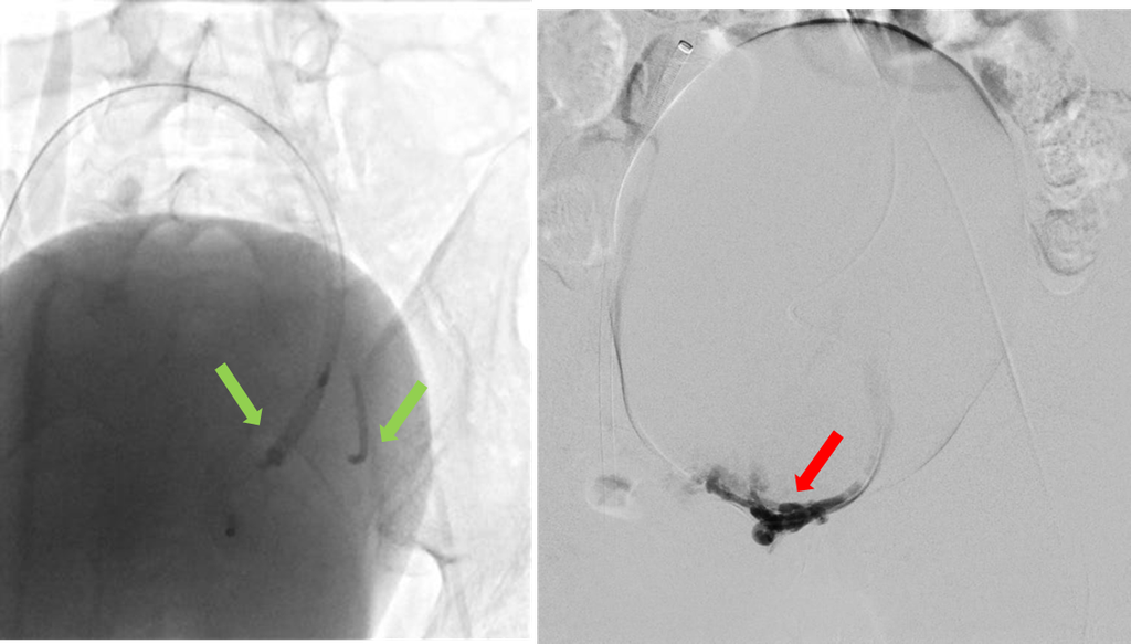
Fig. 2 – Arteriography – left: embolization of the anterior and posterior branches of the left internal iliac vein, using 20 mm and 18 mm plus, respectively (plugs – green arrow); right: main arterial branch of AVM was supra-selectively embolized for nidus embolization with cyanoacrylate:lipidol (1:2) (red arrow).
Post-procedure imaging revealed compression of the S1 nerve root by the posterior plug, causing left-sided sciatica. The patient was treated with prednisone, gabapentin, tramadol, and physiotherapy, resulting in significant pain relief, with pain severity decreasing from 10/10 to 4/10.
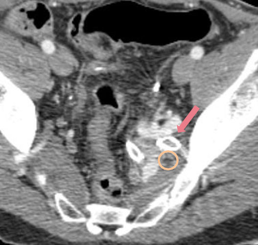
Figure 3 and Video 3– Immediate post-procedural CT revealing compression of the S1 nerve root by the posterior plug. S1nerveroot(circle);posterior plug (pink arrow).
A follow-up CT on May 15, 2025, showed residual perfusion of the AVM and persistent contact between the posterior plug and the S1 root, prompting a second embolization procedure in July 2025.
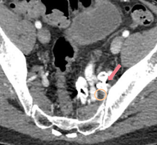
Figure 4 and Video 4 – CT three weeks after procedureprocedure revealing compression of the S1 nerve root by the posterior plug. S1nerveroot(circle);posterior plug (pink arrow).
Treatment Plan
Second embolization procedure involved super-selective catheterization and embolization with microcoils and Onyx, successfully achieving vascular exclusion.
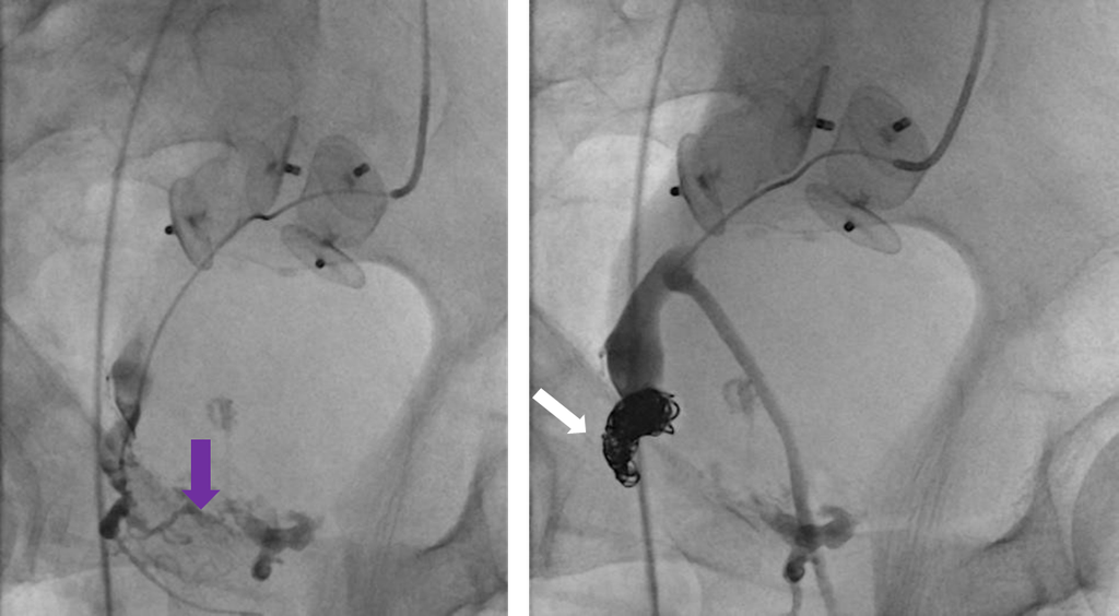
Fig.5— Arteriography. left: angiography of the posterior branch of the left internal iliac artery showing the nidus (purplearrow). right:super-selective embolization of the proximal nidus feeder with microcoils and Onyx (white arrow).
Results and Follow-up
A subsequent CT on August 2025, confirmed thrombosis of the AVM and occlusion of the treated branches, with no significant changes. There is no compression of the S1 nerve root.
The patient showed continued improvement, with resolution of sciatica after discontinuing gabapentin. This case highlights the challenges of managing post-embolization complications in complex pelvic AVMs and emphasizes the importance of ongoing monitoring and tailored interventions.
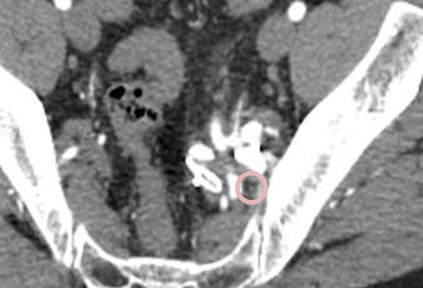
Figure 6 and Video 6– CT one month post-procedure revealed thrombosis of the AVM and occlusion of the treated branches. There is no compression of the S1 nerve root. S1 nerve root – circle.
Discussion
Most neuropathies after pelvic embolization are ischemic (e.g., after internal iliac artery occlusion).
Here, direct mass-effect from a venous plug against the S1 root was the dominant mechanism—an uncommon but plausible cause of radiculopathy.
What to check before deploying a venous plug? (PLUG mnemonic)
- Position: simulate final Position vs foramina on pre-/intra-op CT/fluoro.
- Look-up Lumbosacral root course (S1 exits ~anterolateral sacrum).
- Understand Utflow: choose the venous segment where device will not impinge the nerve.
- Gauge Girth: oversizing can increase mass-effect; consider coils/liquid alternatives.
Evidence-Based Care
- Transvenous ± transarterial strategies for pelvic AVM show high occlusion and symptom improvement; staging is common.
- Onyx/ethylene-vinyl alcohol is effective for peripheral high-flow AVMs as primary or adjunct therapy.
- Ischemic sciatic neuropathy is described after IIA embolization and should stay in the differential of post-embolization sciatica (even when mass-effect is suspected).
Take Home Messages
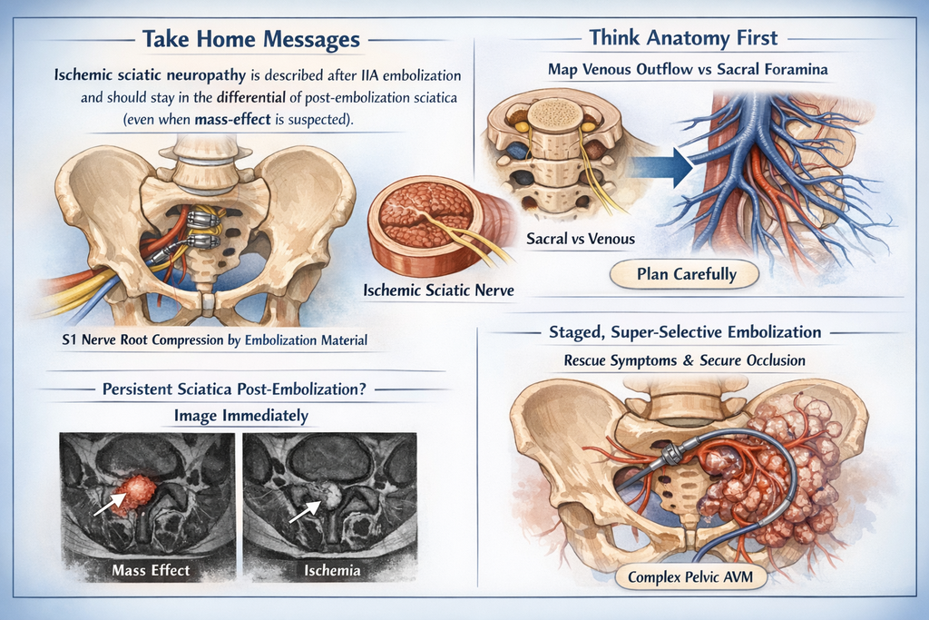
Illustration generated with AI using ChatGPT, based on the author’s prompt; final edits/corrections by the authors.
- Ischemic sciatic neuropathy is described after IIA embolization and should stay in the differential of post-embolization sciatica (even when mass-effect is suspected). S1 nerve root compression by embolization material is a rare complication.
- Think anatomy first: map venous outflow vs sacral foramina when deploying plugs.
- Persistent sciatica post-embolization = image immediately to distinguish mass-effect from ischemia.
- Staged, super-selective embolization can rescue symptoms and secure occlusion in complex pelvic AVMs.
Disclosures
The authors confirm that the case has not been previously published and that informed consent was obtained.